
It is already known that Artificial Intelligence (AI) is a powerful ally in the challenge of increasing companies’ productivity. According to PwC’s Global Artificial Intelligence Study, the estimate is that this technology will increase global GDP by USD 15.7 trillion by 2030.
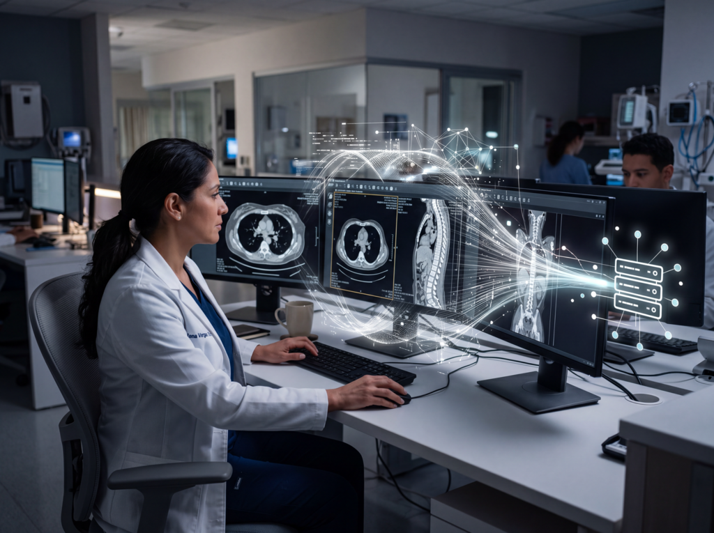
The healthcare field is one of those that benefit from artificial intelligence. AI brings benefits both to institutions and physicians, as well as to patients.
Its use is especially advantageous in radiology and diagnostic imaging clinics that deal with a high volume of requests for exams and reports, and whose application, little by little, is becoming a prerequisite for CDIs that aim to offer a Health 5.0 service.
In this article, understand the role of AI in diagnostic medicine and learn some examples of artificial intelligence for radiology.
The Role of AI in Radiology and Diagnostic Imaging
The main objective of applications with artificial intelligence is to enhance and support human activities, not to replace people.
Therefore, the radiologist’s work is not discarded, only improved. After all, a radiological diagnosis cannot be issued solely based on images.
Regardless of the specialty, the physician needs to evaluate different aspects before preparing a report, such as the patient’s history, symptoms, existing pathologies, genetic and family predispositions, mental health, and even socioeconomic and environmental issues.
AI tools cannot provide or sign diagnoses; rather, they can support professionals in recognizing and differentiating images through suggestions of clinical findings.
In this way, a radiologist needs to assume professional and ethical responsibility for the report, even with this support. Some areas benefit more from advances in technology.
This is the case for complex exams, such as computed tomography (CT), magnetic resonance imaging (MRI), and nuclear imaging. The same applies to organs that are more difficult to diagnose, such as lungs, breasts, and brain, and in certain specialties such as mastology and oncology.
How Does Technology Support Radiologists?
With the use of AI, the expectation is that the radiologist improves time management by shortening manual activities, mainly with regard to quantification.
With this, the professional dedicates more time to analyzing images and the patient’s history, and less to operational activities.
In the past, for example, to detect the coronary artery, it was necessary to perform segmentations manually. Today, the software does this—it segments, characterizes atherosclerotic plaques, and even helps assign the stenosis grading.
In addition, AI helps productivity and the quality of institutions’ professionals’ work.
Some AI algorithms often manage to quantify as well as, or even better than, the radiologist themselves. The variability of measurements performed by algorithms tends to be lower and, therefore, they ensure greater precision.
Main Advantages of Using Artificial Intelligence Solutions in Radiology and Diagnostic Imaging
AI tools have been part of Radiology for quite some time, such as in bone densitometry analysis, voice recognition for preparing reports, and automatic segmentation of coronary arteries.
There were significant gains when they were implemented, and now they go unnoticed because they are incorporated into our day-to-day.
More modern AI tools, with a major impact on radiological productivity, are still in the validation phase. Some benefits are:
- Increased productivity and improved radiology management: by removing operational activities from the radiologist, Artificial Intelligence allows these professionals to save time and to analyze more exams without losing diagnostic precision;
- Agility in diagnosing urgent cases: AI helps prioritize critical findings and prevents the progression of serious diseases. An example is pulmonary thromboembolism and intracranial bleeding, where the tool automatically alerts the radiologist;
- Greater diagnostic safety: the image databases used in this type of tool are updated frequently and contain an incomparably greater amount of content than radiology books and manuals, increasing the reliability of results.
AI Applications for Radiology
The development of AI solutions is based on different techniques and concepts, such as computer vision and deep learning—a method based on artificial neural networks that simulates the behavior of the human brain at a more advanced level.
Currently, radiology software with AI is already capable of identifying uncommon characteristics in medical images, suggesting possible clinical findings that should be evaluated by the radiologist.
In practice, these innovations create an advanced image-recognition capability, based on processing large, up-to-date databases.
Check out some current applications:
AI Helps Detect Rare Cases and Disease Risks
Certain pathologies are difficult to detect, as is the risk of a person developing a given disease. AI solutions include resources capable of assisting physicians in detecting rare cases and particularities.
In addition, when possible diagnoses present very similar images or extreme variability, AI provides greater confidence to the professional when issuing a report.
Cardiovascular Disease Risk Detected Through Retinal Analysis
In 2018, Google Brain, Google’s artificial intelligence research division, developed an ocular scanning program with a specific algorithm to identify retinal data used to detect cardiovascular disease risks, such as age, sex, smoking habits, and systolic blood pressure.
According to data released by the organization, the tool recorded a 70% success rate in tests of images of real people who could have a heart attack or stroke in the next five years.
Currently, retinal analysis is performed only by ophthalmologists. With improved AI tools and applications, this method could become more accessible and practical.
For example, an institution with a retinoscope but no specialist to interpret images—especially for normality triage—could benefit from AI integration.
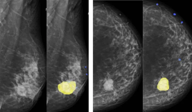
AI for Mammography
Breast cancer analysis involves inspecting mammograms to detect lesions and tumors.
With specialized resources for this exam modality, artificial intelligence can perform the automatic segmentation of breast masses, showing the constant evolution in mammography analyses and suggesting breast findings with density classification and lesion malignancy (BI-RADS). Resources like this can also help with screening, indicating priority of care.
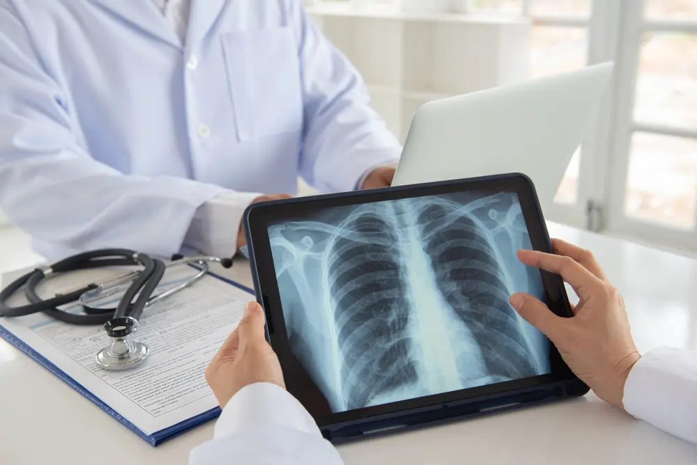
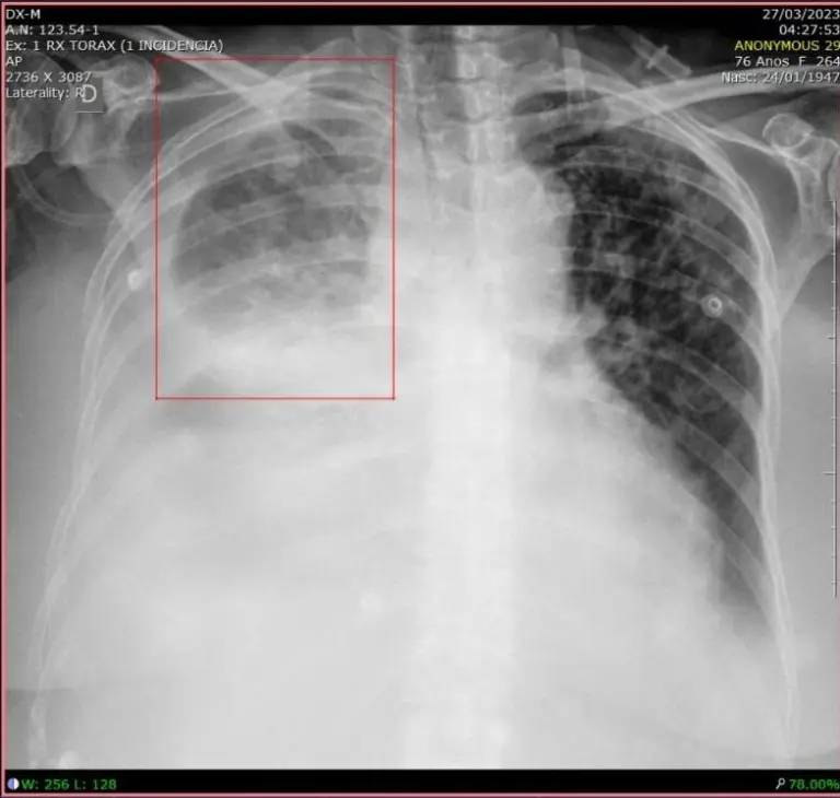
AI for Pulmonary X-Rays
Pneumonia is a lung infection responsible for over 600,000 hospitalizations annually in Brazil’s Unified Pneumonia is a pulmonary infection responsible for more than 600 thousand hospitalizations per year in the Brazilian Sistema Único de Saúde (SUS).
Diagnosing pneumonia on a chest X-ray involves trained specialists and cross-referencing information from clinical history, vital signs, and laboratory tests.
To help with this challenge, machine learning algorithms can detect areas of pulmonary opacity (pleural effusion, cardiomegaly, acute pulmonary edema, pulmonary nodules) and mark them in boxes for medical evaluation, specifying the location and size of any detected infection.
This brings productivity for physicians regarding treatment decisions (mild pneumonia versus severe pneumonia, for example) and also feeds the system back as physicians validate or discard the clinical finding indicated by the computer.
AI for Bone Age Calculation in Hand and Wrist X-Rays
Tools with this focus are capable of estimating an individual’s bone maturity based on the dimensions shown in the hand and wrist X-ray image.
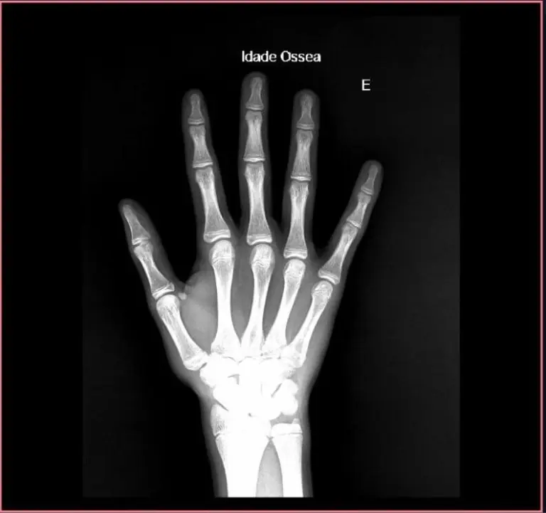
Bone age analysis is especially relevant in areas related to pediatrics. In this case, the calculation algorithm is most relevant as a support basis for the diagnosis of the responsible physician.
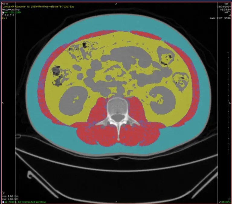
AI for Sarcopenia Calculation
Sarcopenia is defined as a change in skeletal musculature characterized by a reduction in strength and muscle mass secondary to aging, which compromises the individual’s physical performance.
AI tools with this specialty can flag clinical findings and perform an automated measurement of abdominal fat distribution, accompanied by an assessment of muscle mass.
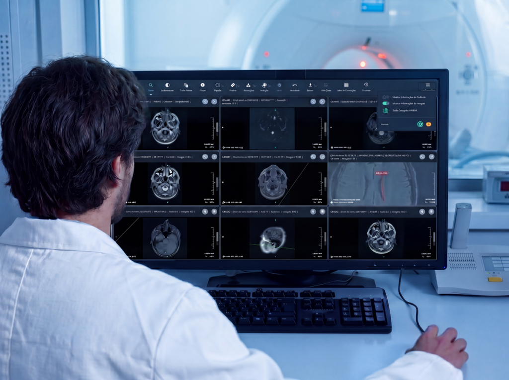
Automatic Vertebra Marker with AI for Radiology
Another possible application is the automatic marker of human and veterinary vertebrae. This is a feature of Pixeon Aurora PACS that helps with locating, marking, and visualizing vertebrae in the sagittal, axial, and coronal planes.
The professional only needs to make the first marking, and the tool performs the remaining ones automatically.
With this, it is possible to increase productivity and precision in CT and MRI exams. See how this feature works:
These are just a few reasons why Pixeon PACS has been chosen four times as the best in Latin America by the U.S.-based Klas Research Institute.
About Pixeon
Pixeon is the company with the largest software portfolio for the healthcare market.
Our solutions serve hospitals, clinics, laboratories, and imaging diagnostic centers, both in management (HIS, CIS, RIS, and LIS) and diagnostic processes (PACS and Laboratory Interface), ensuring higher performance and high-level management in healthcare institutions.
The HIS/CIS software for hospitals and clinics, Pixeon Smart, is comprehensive and integrates the entire institution into a single system, in addition to being certified with the highest level of digital maturity by SBIS (Brazilian Society of Health Informatics).
We already have more than 3,000 clients in Brazil, Argentina, Uruguay, and Colombia, serving millions of patients annually through our platforms.